
We use cookies on this website. Cookies help us deliver the best experience on our website. More about cookies
-
- Education
- Education
- Find programmes and courses
- Applications and admissions
- Tuition fees
- Scholarships
- Exchange studies at Malmö University
- Study Guidance
-
- After admission
- After admission
- Moving to Malmö
- Pre-orientation
- Arrival guide
-
- About studies at Malmö University
- Why choose Malmö University
- Understanding university studies
- Connect with our students
On the page -
- Research
- Research
- Find researchers and research
-
- Doctoral studies
- Doctoral studies
- Doctoral courses
-
- Doctoral schools
- Doctoral schools
- Adaptation of urban space through sustainable regeneration
- Culturally Empowering Education through Language and Literature
- Doctoral school: DACHE
- Education, Learning and Globalisation
- Finding ways in a time of great future challenges (FinnFram)
- Swedish National Graduate School in Science and Technology Education Research
- Learning in Multicultural Societal Contexts
- Pedagogy and Vocational Skills
- Relevancing Mathematics and Science Education (RelMaS)
- Sustainable Movement Education
- The National Research School for Professionals in Social Services
- Research subjects
-
- Research centres
- Research centres
-
- Biofilms Research Centre for Biointerfaces
- Water Science Lab
-
- Citizen Health
- Citizen Health
- Kids in Action
- Imagining and Co-Creating Futures
- Institute for Urban Research
- Malmö Institute for Migration Studies
- Literacy and Inclusive Teaching
- Centre for Work Life Studies
- Sustainable Digitalisation Research Centre
- Centre for Sexology and Sexuality Studies
-
- Research publications
- Search for research publications in Diva
- Malmö University Press
-
- Research in focus
- Research in focus
- Focus: In the classroom
- Focus: The Election 2026
- Focus: A future with AI
- Focus: Climate and society
- Focus: Peace, democracy and human rights
- Focus: The mouth
- Focus: Migration
- Focus: Elite Sport, Football and Supporter Culture
- Focus: Perspectives on Malmö
- Focus: Loneliness
- Research events
- Participate in a research study
On the page -
- Collaboration & Innovation
-
- Levels of collaboration
-
- Local collaboration
- Muvah
- Co-Create Malmö
- Regional collaboration
- National collaboration
-
- International collaboration
- UNIC
- Innovation
- Collaboration with students
- Collaborate with researchers
- Culture collaboration
- Support Malmö University
On the page -
- About us
- About us
-
- Meet Malmö University
- The history of the University
- Malmö University in numbers
- Sustainability
-
- Management and organisation
-
- Faculty of Culture and Society
- Department of Global Political Studies
- School of Arts and Communication
- Department of Urban Studies
-
- Faculty of Education and Society
- Department of Childhood, Education and Society
- Department of Sport Sciences
- Department of Culture, Languages and Media
- Department of Natural Science, Mathematics and Society
- Department of Society, Culture and Identity
- Department of School Development and Leadership
- The Centre for Teaching and Learning (CAKL)
- Knowledge Centre for Educational Health in Schools (PHIS)
-
- Faculty of Technology and Society
- Department of Computer Science and Media Technology
- Department of Materials Science and Applied Mathematics
- Faculty of Odontology
- Management and decision-making paths
- Malmö University's strategy 2030
-
- Quality assurance work at the University
- Widened recruitment and participation
- University Dental Clinic
-
- Malmö Academic Choir and Orchestra
- Student work – video pieces
On the page -
- Contact
- Contact
- We help you with your questions
- Find your way around Malmö University
- Visit Malmö University
-
- News and press
- News and press
-
- Graphic manual
- Graphic manual
- Logo
- Colours
- Typography
- Visual language
- Film and live broadcasts
- Merchandise
- Supplier information and invoice management
- Whistleblowing
On the page
Imaging
infrastructure
Advanced imaging facilities for research and collaboration. We offer advanced imaging technologies for studying biological samples, materials and structures, from the nanoscale to larger complex systems. The facility provides access to specialised equipment, expert support and image analysis for both internal and external researchers.
Available equipment
Three principally different types of microscopes are available:
- Light microscopy – for lower-resolution imaging of reflected or transmitted light,
- Confocal laser scanning microscopy – for higher-resolution imaging of transmitted light, including 3D imaging, but only for fluorescently labelled or auto-fluorescing samples,
- Scanning electron microscopy – for highest-resolution imaging of reflected light (electrons).
Light microscope
The Nikon Eclipse LV100ND is an upright research-type light microscope with LED illumination, capable of imaging both transmitted and reflected light. The dual light source setup allows the microscope to be used both for transparent samples, such as fixed biological samples or lipids (transmitted light), as well as for material science applications such as metals or polymers (non-transparent samples, reflected light).
More about Nikon Eclipse LV100ND light microscope
- The microscope is equipped with a Linkam Analysa-LTS350 temperature-controlled stage to allow imaging of samples at elevated temperatures, for example to investigate temperature-induced phase transitions. It is further equipped with polarising filters for both reflected and transmitted light, to allow study of birefringent samples. There are two sets of objectives, one for short working distance (10X, 40X, 100X) for imaging of flat samples with cover glass; and one set with long working distance (10X, 50X), for imaging the surface of non-flat samples or when using the temperature-controlled stage. A TransferMan NK2 micromanipulator allows manipulation of samples during imaging, such as mounting of AFM colloidal probes. The microscope is equipped with a CAM-UC90, 9 megapixel, 1” CCD, 4k resolution digital camera and is running cellSens Entry V2.
Specifications
- Upright light microscope
- LED illumination for both transmitted and reflected light
Available objectives:
- CFI Plan Akromat 10X, N.A. 0.25, W.D. 10.5 mm
- CFI Plan Akromat 40X, N.A. 0.65, W.D. 0.56 mm
- CFI Plan Akromat 100X olja, N.A. 1.25, W.D. 0.2 mm
- CFI TU Plan Fluor Epi 10X, N.A. 0.30, W.D. 17.5 mm
- CFI TU Plan Epi ELWD 50X, N.A. 0.60, W.D. 11 mm
Special features:
- Temperature-controlled stage Linkam Analysa-LTS350
- TransferMan NK2 micromanipulator
- Polarising filters for both reflected and transmitted light
- Acquisition Software: cellSens Entry V2

Confocal laser scanning microscope
The Evident FLUOVIEW FV5000 is a fully motorised, inverted, high-performance laser scanning confocal microscope designed for advanced biomedical research. The system enables high-resolution, quantitative, and three-dimensional fluorescence imaging of fixed and live specimens. The microscope can be used to visualise cellular and subcellular structures, monitor dynamic biological processes, and analyse complex tissues with high spatial and temporal resolution. Applications include multiplex fluorescence imaging with up to six channels simultaneously, imaging of thick tissue and organoids, and quantitative analysis.
More about Evident FLUOVIEW FV5000 confocal laser scanning microscope
The system also supports live-cell imaging, as it is equipped with a temperature- and CO2-controlled climate chamber. In addition to the galvanometric scanner, the platform includes a resonant scanner that provides ultra-fast image acquisition, making it well-suited for imaging dynamic biological processes. The instrument can be used in research areas such as cell biology, neuroscience, developmental biology, cancer research, immunology, and drug discovery. The microscope supports studies ranging from single-cell analyses to large-scale tissue imaging, and facilitates investigations of molecular localisation, cell-cell interactions, and physiological processes.
Specifications
Laser lines: 405 nm, 445 nm, 488 nm, 561 nm, 594 nm, 640 nm
Detectors:
- 4 low-noise SilVIR type detectors (2 blue sensitive, 2 red sensitive)
- 1 transmitted light detector
Scanners:
- 8kx8k Galvanometric scanner, for best quality images
- 2kx2k Resonance scanner, for fast imaging
Available objectives:
- UPLXAPO 4X NA 0.16 (dry)
- UPLXAPO10X O NA 0.40 WD 3.1mm (dry)
- UPLXAPO 20X NA 0.80 WD 0.6mm (dry)
- UPLAPO 25x NA 1.0 WD 1.0mm
- UPLXAPO 40x NA0.95 WD0.18 (dry)
- UPLSAPO 60xOHR NA1.50 WD 0.11 (oil, highest numerical aperture)
- PLAPON 60XOSC2 NA 1.40 WD 0.12 (oil, super-corrected, for co-localisation studies)
Acquisition Software: cellSensFV
Special Features:
- Stage-top climate control chamber Okolab UNO+
- LED fluorescence light source for transmitted and reflected light
- High-resolution camera for wide-field fluorescence imaging or non-fluorescent staining
- Software-controlled auto-correction collar for one objective
- Auto-adjusted laser intensity
- Auto sample finder
- Auto-focus finder with hardware-based z-drift compensator
- AI-supported de-noising functions
- Live and post-processing deconvolution functions
- FV-OSR Super resolution module for X-Y imaging down to 120 nm

Scanning Electron Microscopy (SEM)
Zeiss EVO LS-10 scanning electron microscope. A scanning electron microscope uses an electron beam that sweeps over the sample pixel by pixel, and by collecting emitted or reflected electrons from the sample, it can create an image of the sample surface. Due to the shorter wavelength of electrons compared to light, maximum resolution is higher, in principle down to 2 nm for the EVO LS-10. It allows imaging of samples over a wide range of magnifications (the EVO LS-10 can, for normal working distances, give a sharp picture at any magnification in the range 50X-50000X) and with a much larger focal depth than a light microscope. This is especially important for non-flat samples, where one wants the entire view to be in focus even though different parts are at different heights.
More about Zeiss EVO LS-10 scanning electron microscope
By using different types of detectors, different types of signals can be collected, revealing complementary information about the sample. The standard secondary electron detector will give the best signal-to-noise and thereby the best image quality. The backscatter detector detects only high-energy backscatter electrons, potentially originating from deeper into the sample, which gives a flatter and less detailed image, but additionally offers information about sample composition, as differences in intensity will indicate differences in atomic weight of the exited elements. By collecting X-ray radiation generated by the sample, the EDX (electro-dispersive X-ray) detector can identify and quantify the relative composition of the elements in selected parts of the sample, either for larger areas (entire part of the sample currently imaged), point and ID (for selected parts/features of the sample) or by mapping.
The Zeiss EVO LS10 is an environmental scanning electron microscope, equipped with a LaB6 filament. The EVO LS10 offers two additional imaging modes, apart from the standard high vacuum mode for fully conducting samples. In variable pressure mode, a low pressure of nitrogen gas (usually 10-40 Pa) is allowed in the chamber. The gas is ionised by the electron beam and the ions help dissipate the surface charge built up in poorly conducting samples, allowing imaging of non-conductive samples without surface coating. In extended pressure mode, water vapour is introduced into the chamber, which, together with a Peltier Coolstage, allows imaging of hydrated samples at controlled temperature and relative humidity.
An Agar automatic sputter coater is also available for gold coating of samples.
Specifications
- LaB6 filament, 1-30 kV
Detectors:
- Everhart-Thornley Secondary electron detector
- Lens mounted 4Q-BSD backscatter detector
- VPSE G3 (Variable pressure secondary electron detector)
- INCA x-act LN3-free EDX detector
Imaging modes:
High vacuum
- Best image quality, for conductive or gold-coated samples
Variable pressure
- Lower image quality, for non-conductive samples where coating is impossible or unwanted
- 10-40 Pa of nitrogen gas in the sample chamber
Extended pressure
- For cooled, hydrated samples such as cells/bacteria, skin, polymers
- Temperature-controlled Peltier stage
- 1-3000 Pa of water vapour in the sample chamber, allows controlled relative humidity
Sample holder and stage:
- 8-position sample carousel for Zeiss 13 mm pin-type sample stubs
- Multi-purpose quick-fit sample holder for larger irregular specimens
- Maximum specimen height 100 mm, maximum diameter 230 mm
- 5-axis stage: X, Y, Z, rotation (0-360) and tilt (0-90)
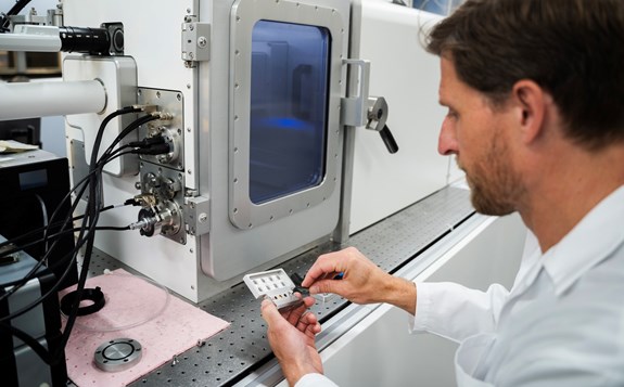
Imaging infrastructure in short
What is it?
A multidisciplinary imaging infrastructure providing access to advanced microscopy technologies, including light microscopy, confocal laser scanning microscopy and scanning electron microscopy.
What is it used for?
To visualise, analyse and characterise biological, biomedical and material samples across multiple length scales, from micrometre-level structures to nanoscale features. The infrastructure supports both routine imaging and advanced applications such as 3D imaging, live-cell studies, surface characterisation and elemental analysis.
Why is it important?
Different research questions require different imaging approaches. By combining complementary microscopy techniques within a single infrastructure, researchers can gain detailed insights into the structure, composition and behaviour of samples, enabling discoveries across life sciences, health research and materials science.