Laboratories and equipment
Research resources
The University’s research infrastructure includes, amongst other things, laboratories for chemistry, cell and microbiological studies, clinical measurement systems, advanced imaging equipment and analytical platforms. Some of these research resources are presented here.
Examples of research resources


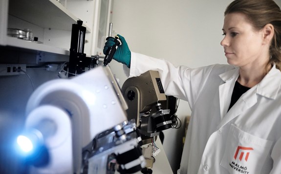
Open lab Skåne
At Open Lab Skåne, you will find equipment for studies ranging from whole tissue to nanoscale and expertise in materials science, physical and colloid chemistry, analytical and synthetic chemistry, biochemistry, and cell and molecular biology. We also support companies interested in neutron and X-ray scattering techniques.
The mission of Open Lab Skåne is to facilitate innovation by opening up our laboratories and enabling access to equipment and expertise. The project is funded by the European Regional Development Fund and Region Skåne. In addition, Open Lab Skåne is supported by Medeon, Medicon Village, CLUK, the city of Malmö, the city of Lund, and the Sten K. Johnson Foundation.
Research in action
Bone healing with metals that gradually dissolve
Synchrotron X-ray tomography for non-destructive 3D imaging
Synchrotron-based X-ray tomography is a powerful and non-destructive imaging method with a wide range of applications that span biomedicine, materials science, engineering and nanotechnology; it allows measurements of the internal structure of materials in 3D at high speed. At Malmö University, the technique has been utilised to study bone growth and healing, the studies of wood coatings, and the penetration of drugs and cosmetics through the skin.
The challenge of permanent metal implants
One of the most common surgeries in orthopaedics is the internal fixation of fractures with metal implants. This includes pins, plates, screws and rods, which are used to reconnect and hold together the fractured parts and allow them to heal. Implants are also used for bone reconstruction in cranial and facial surgery. The most used and well-functioning materials are titanium alloys and stainless steel. A major drawback with these materials is the need for their removal after the bone heals. For the patient, this often involves a second surgery, with all the risks and complications associated with it. Designing self-degrading implants that can fixate fractures and then disappear by themselves when the bone is healed is therefore of significant interest.
Magnesium as a biodegradable implant material
Magnesium is a metal that is naturally present in the body and has strength similar to that of the bone. It also has the property of gradually dissolving in the body to form non-toxic products. It shows great promise as an implant solution for use in load-bearing areas of the skeleton. In fact, the constant release of Mg ions in the fracture site could potentially have a positive stimulus for bone growth and magnesium-based implants were therefore produced and tested as bone implants.
Tracking implant degradation over time
Through the use of synchrotron X-rays, images can be produced at a rapid rate, allowing scientists to follow implant degradation in a time-lapse manner.
I can't believe it's butter
Understanding fat crystal structures
Fat crystallography is very important for numerous applications in industries spanning food, cosmetics and pharmaceuticals. Fats' rheological and mechanical properties depend largely on the structure of the fat crystal networks. The increasing interest in lipid excipients for the development of taste-masked and modified drug release formulations is often compromised by release instability. Further mechanistic understanding in this regard is also required to facilitate larger-scale applications.
The mystery of the Vasa butter
Sweden’s most famous battleship, the Vasa, was floated in 1628 and salvaged from the seabed with a nearly intact hull in 1961. Archaeologists were faced with a largely undisturbed 4-storey structure to excavate. Among the findings, on board were various containers with butter. The butter was found to be very salty and had apparently turned rancid from 333 years of storage at four degrees centigrade. A sample from a smaller tin canister was later analysed by Malmö University researchers.
Investigating centuries-old fat degradation
In 1991, structural investigations were carried out using the X-ray equipment available to the researchers at that time. Complementary chemical analysis confirmed that a large part of the main constituent in the butter, the triglycerides, had degraded to free fatty acids (mono- and diglycerides). The total fatty acid composition, however, deviated from that of fresh butter, and they came to question whether the tin canisters had really been watertight or if some of the content had dissolved into the seawater.
New opportunities with synchrotron X-rays
The evolution of new generations of X-ray equipment has provided us with much more powerful tools than the ones we used for the Vasa study. Access to synchrotron light at MAX IV can also be combined online with other complementary techniques.
Capturing phase transitions in real time
The high-energy X-rays of the synchrotron permit the recording of powder diffraction patterns in as little as two seconds, meaning that thermally induced changes can be accurately quantified and additional insight into the nature of phase transitions can be obtained. Such detailed knowledge cannot be obtained from existing laboratory instruments, which require a much longer collection time.
Good and bad cholesterol
The hidden processes behind heart disease
Atherosclerosis, where plaques of fats and cholesterol build up in the arteries, can lead to heart attacks and strokes. Certain lipoproteins, the ‘good’ and ‘bad’ cholesterol, function as cholesterol and fat transporters in the blood and are currently used as clinical markers for disease. Measuring the total levels of cholesterol or lipoprotein particles does not accurately predict the risks of developing atherosclerosis, as many people suffer from cardiovascular disease even though they show normal lipid clinical profiles.
Following the interaction between blood fats and artery walls
In order to understand the molecular mechanisms behind plaque build-up, it is necessary to understand both the lipoprotein particle structure and the structure of the artery wall during their interplay. However, both these systems are complex molecular assemblies and determining their detailed structure has proved to be extremely challenging.
Seeing what other techniques cannot
One of the main advantages of neutrons is that they see hydrogen and deuterium atoms differently. This in turn allows for the highlighting of different parts of a complex biological system individually.
From fundamental research to future treatments
For interactions between native lipoproteins and the arterial wall, neutron scattering in combination with selective deuteration has provided an elegant way to systematically study each component individually. Such knowledge will not only aid in improving current diagnostic tools, but could lead to the development of more specific drugs.
Neutrons help understand nature’s own lubricant
Looking beneath the surface
Neutron reflection is a powerful technique employed to see deep into the matter and to deliver profound information on surfaces. By developing a new instrument that can provide a way to study the lubrication of thin films under both confinement and shear, we can perform neutron reflectometry studies and a technique known as GI-SANS studies. These can provide in-plane information near the surface of thin films.
Learning from nature's lubricants
Throughout history, we have been using oil-based lubricants to help prevent damage and tear. Our own body, however, has developed water-based systems to grease its sheared surfaces. These bio-lubricants, such as saliva and mucus, outperform any man-made oil-based greasing agents. But why are they so extremely difficult to imitate? Understanding how nature overcomes the drawbacks of water as a lubricant is one of the projects at Malmö University that can lead to the design of novel lubricating formulations.
Understanding how lubrication works
Shear is expected to initiate changes in the structure of thin films, and the characterisation of these changes is critical for understanding their lubrication mechanisms. This, on the other hand, is essential for the prediction of the lubricating performance of thin films based on their composition and therefore the rational development of novel lubricating formulations.
From fundamental insights to real-world applications
The methodology that enables studies of the bio-lubricants found in our body can give fundamental knowledge of their structural and mechanical characteristics. This can lead to the design of non-toxic, aqueous lubricating systems with novel biomedical applications and help replace the highly contaminated oil-based lubricants currently used in our engineering practices. It will also allow for the development of new treatments for several diseases that are associated with changes in our bio-lubricating properties.
Skin – the final frontier
Advancing topical treatments
Extensive research has been carried out at Malmö University into the development of non-invasive or minimally invasive topical applications — whether that be for medical or cosmetic purposes. Collaborating with other universities and industry partners, this has involved utilising the MAX IV facilities.
How the skin adapts to its environment
Skin responds to changes in the environment, for example, change from dry skin in a cold and dry climate to moist, wrinkly skin after a long hot bath, or from the application of a cosmetic cream to alleviate dry skin conditions. From a scientific perspective, it is relevant to understand how and why the skin barrier is affected by changes in the surrounding conditions and how this influences the function of the skin barrier. This can in turn help us understand the uptake of drugs applied to the skin surface, progression of skin disorders and skin health in general.
Revealing the skin barrier at the molecular level
X-ray diffraction is a compelling technique used to investigate the structural features of the skin barrier. In our research, understanding how this structure-function relationship is influenced by parameters such as relative humidity, temperature, or application of common substances that are used in commercial skin care products is of particular relevance.
Thin films are all around us
The hidden structure of thin films
Thin polymeric films have numerous applications in a variety of fields, such as packaging, membranes, paints and coatings, adhesives, electronics and textiles, as well as cosmetics and biomedical devices. They can be prepared from a wide range of different formulations. Of specific interest to Malmö University researchers are latex films. For the understanding of the performance of these films, it is of high importance to characterise the structure of the film’s outer layer.
Following the journey from particles to film
Typical sizes for polymer particles used for waterborne coatings are in the 50-200nm range. Additionally, smaller structures may also be found inside latex films. Small-angle X-ray scattering is a technique that can be used for particles in this size range and was used to probe the merging of polymer particles during the film formation as well as to detect the presence of smaller structures within the films.
The challenge of capturing film formation
One of the weaknesses of standard X-ray scattering experiments is that they require sample dimensions in, at least, the millimetre range. This is above what is relevant for most coating applications and subsequently results in longer drying times and difficulties in monitoring the film formation process. In this regard, a grazing incidence angle geometry stands out as a more suitable technique. This technique, where the incident X-ray beam strikes the sample at a small angle, can be used to provide structural information about nm-thick films. This technique is currently being explored to monitor the formation of latex films in real time.
Understanding superalloys
Advanced alloys for demanding environments
Metallic alloys are a combination of two or more metallic or other elements, such as steel (a combination of iron and carbon) and bronze (a combination of copper and tin). A typical family of alloys — that has superior properties in comparison with other alloys — are the so-called super or high-performance alloys. These alloys are characterised by excellent mechanical properties and can resist high mechanical loads at elevated temperatures. A typical application for such alloys is in turbine engines, heat exchangers, and power plants.
Understanding what makes superalloys perform
From a standard tensile test, the mechanical properties for an alloy can be measured at different temperatures and for different conditions, such as solution-treated or age heat treated. This can provide information on the precipitates' impact on the mechanical properties of the alloy. However, this method does not directly measure the precipitate size, the volume fraction and how they develop during a heat treatment, which is important to understand when developing new heat treatments and for developing new superior superalloys.
Following changes inside the material
High-energy synchrotron X-rays enable in-situ measurements of precipitation in alloys at different thermal and mechanical loadings. This can provide information on the particle size distribution, the volume fraction of the precipitates and their shape.
Towards stronger and more energy-efficient materials
A compact instrument able to measure the response of the precipitates during mechanical loading has been developed through a joint project which included academics from Malmö University. The advancement of the technique enables scientists to understand the mechanism of precipitation and how they interact with the alloy, which can eventually lead to new heat treatments that can save energy.